S D S Cardiac Anatomy

This category has only the following subcategory.
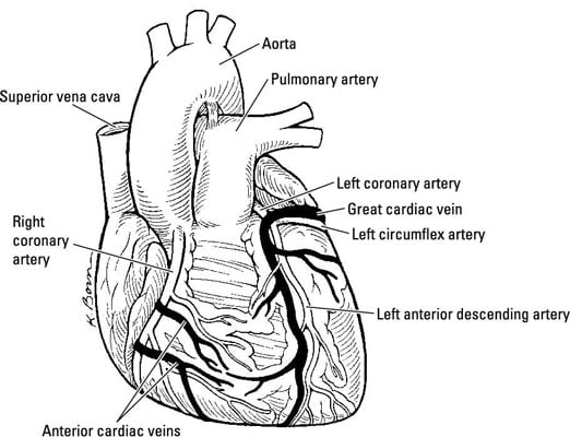
S d s cardiac anatomy. Structure of the heart the heart is a muscle about the size of a fist and is roughly cone shaped. It is about 12cm long 9cm across the broadest point and about 6cm thick. A bundle of fibers that carry cardiac impulses. What is the heart.
Although the ventricles on the right and left sides pump the same amount of blood per contraction the muscle of the left ventricle is much thicker and better developed than that of the right ventricle. It is enclosed in the mediastinal cavity of the thorax between the lungs and extends downwards on the left between the second and fifth intercostal space fig 1. A section of nodal tissue that delays and relays cardiac impulses. This article describes the heart s anatomy and physiology.
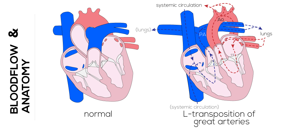
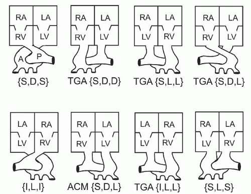
The great vessels are said to be inverted with the aortic valve to the left relative to the pulmonary valve and lie in a parallel rather than crossed position as in the normal anatomy. The heart is located in between the two lungs. Figure 19 7 heart musculature the swirling pattern of cardiac muscle tissue contributes significantly to the heart s ability to pump blood effectively. Although sll is more common than idd it is still considered a rare defect with an incidence of 0 02 0 07 per 1 000 live births.
A few individuals may have both s 3 and s 4 and this combined sound is referred to as s 7. The heart weighs around 350g and is roughly the size of an adult s clenched fist. It lies left of the middle of the chest. Heart nodes and nerve fibers play an important role in causing the heart to contract.
Cardiac anatomy refers to the structure of the heart one of the organs of the body. V heart valves 6 p pages in category cardiac anatomy the following 69 pages are in this category out of 69 total.


